Electrical Impedance Tomography (EIT) is a medical imaging technique employing voltage applied to different points on the body to reconstruct an image of what’s inside.
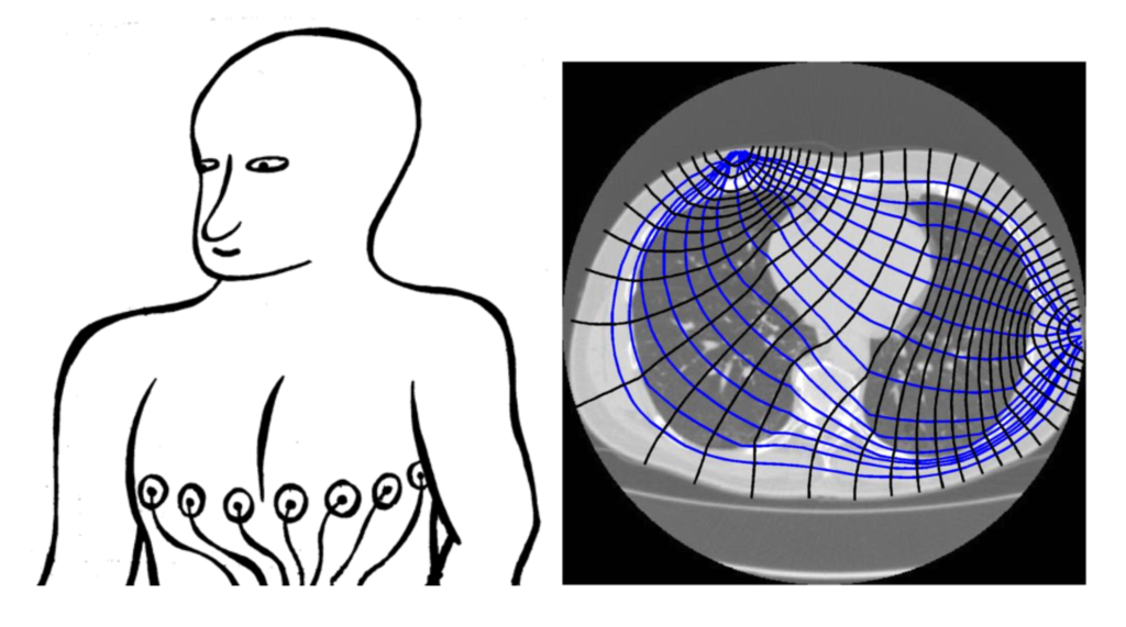
From a mathematical standpoint, EIT is related to the solution of a D-bar problem. From a medical standpoint, EIT has the potential to provide valuable, rapid medical imaging for the diagnosis of conditions such as aneurysms and pulmonary embolisms, but its uses are currently limited due to the low resolution of the images on offer.
I am now managing a project to improve the quality of EIT images through techniques in mathematical physics and mathematical computing.
This poster by IMB postdoctoral fellow Nikola Stoilov provides more information on the project.
Thanks to our funders for their generous support.